
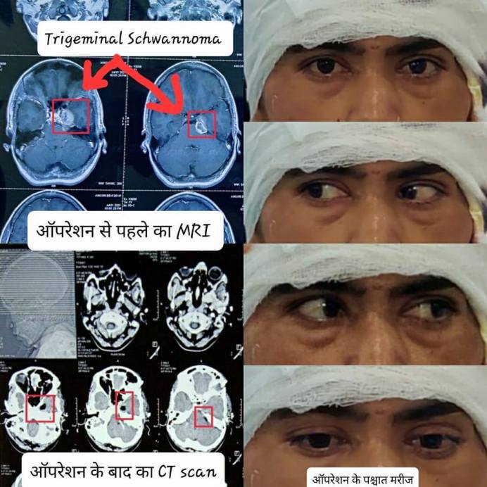
Trigeminal Schwannoma
Procedure Done by : Dr. Abhishek Chauhan & Team
Trigeminal Schwannoma
Did subtemporal interdural approach and excision of tumor (Trigeminal Schwannoma) in a 36 year old lady.
She presented with complaints of left eye deviation and drooping of eyelid since past 10 days. She also had redness of left eye, severe left facial pain, decresed sensation over left cheek and jaw deviation on opening mouth since past 1month. On examination she was found to have partial 3rd and 5th cranial nerve palsy. After surgery her symptoms were relieved as shown in picture and she recovered well.
Trigeminal Schwannoma
They are rare benign tumors originating from the Trigeminal nerve (nerve responsible for jaw movement and facial sensation mainly). It is situated at critical location surrounded with the cranial nerves related to the movement of our eyeball and cavernous sinus. So the surgery in this area is risky and can put patient at risk of losing eye movement, not able to open eye (Ptosis) and severe blood loss. But we were able to remove the tumor preserving surrounding nerves because of the advanced neurosurgical setup we have at BIMR Hospital Gwalior especially
- leica Microscope which magnifies the tumor and surrounding nerves to facilitate safe surgery and
- CUSA (Cavitron UltraSonic Aspirator) which disintegrates tumor with the help of high frequency ultrasound waves and then suck them at the same time without disturbing the surrounding structures.
ट्राइजेमिनल श्वानोमा (Trigeminal Schwannoma)
ये एक दुर्लभ ट्यूमर हैं जो की ट्राइजेमिनल नामक नस जो कि आंख व चेहरे से सिग्नल दिमाग तक पहुंचाने के लिए जिम्मेदार होती है से उत्पन्न होते हैं। यह ट्यूमर केवरनस साइनस व आंख की हरकत के लिए जिम्मेदार नसों से चिपका रहता है जिस कारण सर्जरी काफी जटिल व जोखिम भरी होती है। और ऑपरेशन के बाद रोगी को आंखों की गति खोने, आंख खोलने में सक्षम नहीं होने और गंभीर रक्त हानि के जोखिम में डाल सकता है।
लेकिन Bimr Hospitals gwalior में अत्याधुनिक न्यूरोसर्जरी सेटअप होने के कारण हम आसपास की नसों को बिना छेड़े ट्यूमर को निकालने में सक्षम हो पाए। इन उपकरणों का महत्वपूर्ण योगदान रहा -
- leica माइक्रोस्कोप जो कि ट्यूमर और आसपास की नसों को कई गुना बड़ा दिखाता है जिससे सर्जरी सुरक्षित रूप से हो सके।
- CUSA (कैविट्रॉन अल्ट्रासोनिक एस्पिरेटर) जो उच्च आवृत्ति अल्ट्रासाउंड तरंगों की मदद से ट्यूमर को विघटित करता है और फिर आसपास की संरचनाओं को छेड़े बिना उन्हें एक ही समय में चूस कर निकाल देता है।